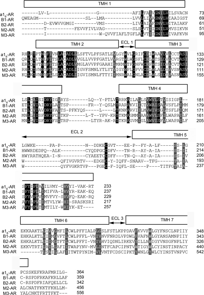
FIGURE 1.
Structural alignments of hamster α1B-AR, human β1-AR (PDB code 2VT4), human β2-AR (PDB code 2RH1), human M2-AR (PDB code 3UON), and rat M3-AR (PDB code 4DAJ). The three-dimensional structural alignments were performed using program TOPOFIT (55) with a joint distance cutoff of 3 Å between the Cα atoms of the residues in the α1B-AR model and each crystal structure. The alignment represents the degree of conservation of residues spatially. The poorly aligned regions indicate residues are not topologically close, although the sequence may be conserved.

