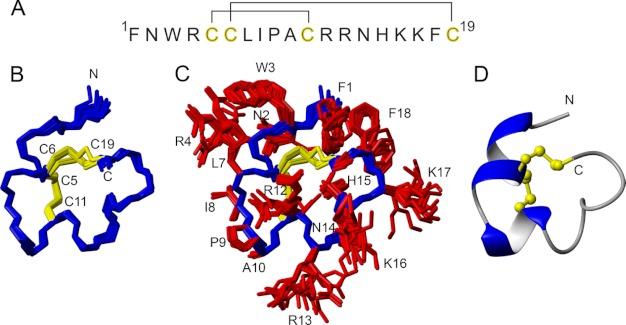
FIGURE 5.
Sequence and structure of ρ-TIA. A, primary sequence and disulfide connectivities. B and C show a superposition of the final 20 structures representing the three-dimensional NMR structure of ρ-TIA (PDB code 2LR9) without and with side chains, respectively. Residues are labeled with single letter amino acid codes and residue numbers. D shows the lowest energy structure in ribbon style, illustrating the elements of secondary structure, and the disulfide bonds in ball-and-stick representation.

