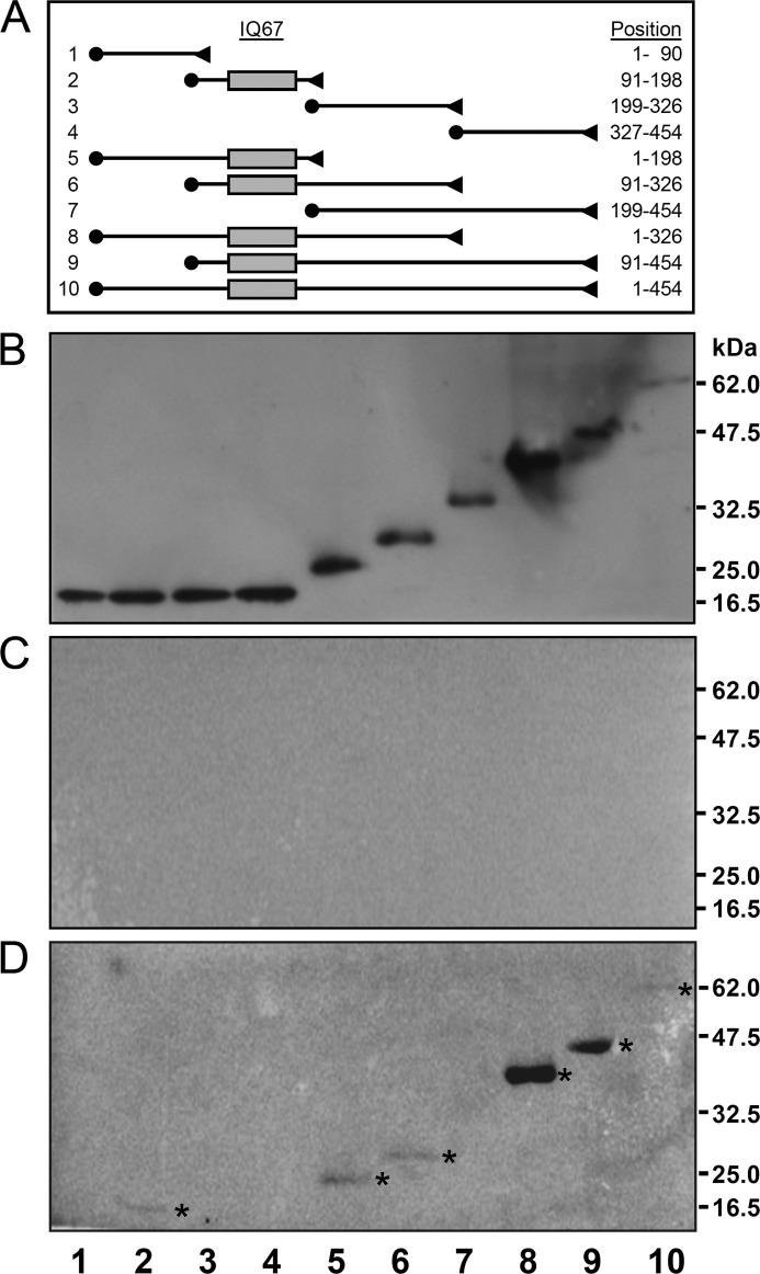
FIGURE 2.
Mapping of the CaM binding domain in IQD1. Full-length epitope-tagged T7-IQD1-His6 and nine derived truncated IQD1 polypeptides were purified by affinity chromatography on Ni-NTA, separated by SDS-PAGE, transferred to a membrane, and probed with [35S]Met-labeled CaM2 in the presence of 1 mm CaCl2 or 5 mm EGTA as described under “Experimental Procedures.” A, shown is a map of the 10 epitope-tagged IQD1constructs. The N-terminal T7 tag and the C-terminal His6 tag are indicated by solid circles and triangles, respectively. The gray box denotes the IQ67 domain, and the length of each IQD1-derived polypeptide is given on the right (position of amino acid residues). B, shown is immunodetection of SDS-PAGE, resolved Ni-NTA-affinity-purified IQD1 polypeptides with an HRP-conjugated T7-tag monoclonal antibody. C and D, overlay assays with [35S]Met-labeled CaM2 in the presence of 1 mm CaCl2 (C) or 5 mm EGTA (D) is shown. Note only IQD1-derived polypeptides containing the IQ67 domain bind to Arabidopsis CaM2 (asterisks in D).

