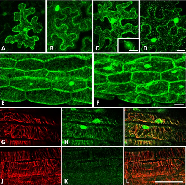
FIGURE 6.
GFP-tagged IQD1 proteins localize to microtubules and the cell nucleus. A–D, tobacco leaves (N. benthamiana) were transiently transfected with Agrobacterium strains harboring plasmids supporting expression of CaMV 35SPro::GFP (A), CaMV 35SPro::IQD1∼GFP (B), and CaMV 35SPro::GFP∼IQD1 (C and D). Samples were collected 2 days after infiltration and visualized by confocal laser scanning microscopy after mock treatment with DMSO (A–C) or after infiltration with 50 μm oryzalin for 90 min (D). E and F, transgenic Arabidopsis seedlings (CaMV 35SPro::GFP∼IQD1) were visualized (three-dimensional projections) before (E) or after (F) treatment with 5 μm oryzalin for 60 min. G–L, GFP∼IQD1 and microtubules were co-immunolabeled in transgenic (G–I) and wild type (J–L) Arabidopsis seedlings using a combination of a rat anti-α tubulin antibody (G and J) and a green fluorescent monoclonal mouse anti-GFP antibody (H and K). Merged images are shown (I and L). Scale bars, 20 μm. Nuclear localization of GFP-tagged IQD1 was additionally demonstrated by DAPI (4′,6′ diamino-2-phenylindole·2HCl) staining (see supplemental Fig. S4).

