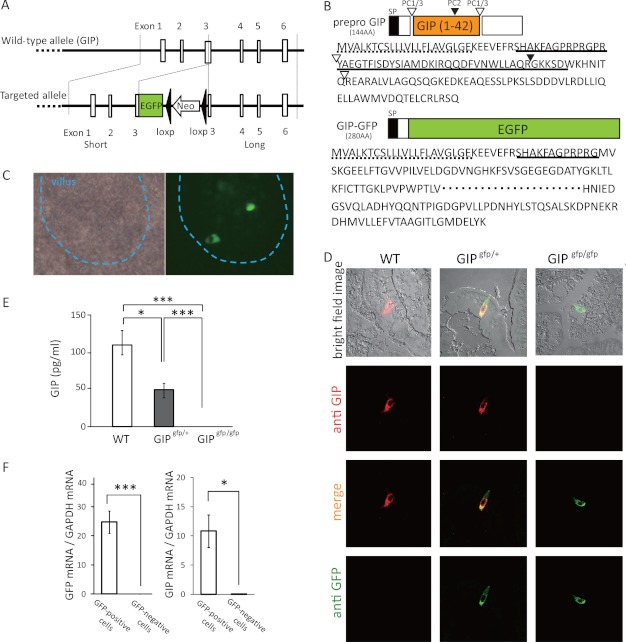
FIGURE 1.
Gene construct of GIP-GFP mice. A, wild-type GIP allele and targeted allele of GIP-GFP. EGFP-poly(A)-loxp-Neo-loxp cassette was inserted into exon 3 of wild-type gip gene. B, prepro-GIP protein and GIP-GFP fusion protein. SP, signal peptide. Open triangle, PC1/3 cleavage site; closed triangle, PC2 cleavage site; dotted line, amino acids of signal peptide; solid line, translated protein from exon 3. C, microscopic images of upper small intestine in GIP-GFP heterozygous mice (bright field image and fluorescence image). D, immunohistochemical images of upper small intestine in wild-type (WT), GIP-GFP heterozygous (GIPgfp/+), and homozygous mice (GIPgfp/gfp). Green, GFP-expressing cells; red, GIP-expressing cells; yellow, merged image. E, fasting plasma GIP levels in WT, GIPgfp/+, and GIPgfp/gfp mice. F, GFP mRNA and GIP mRNA levels in GFP-positive cells (n = 5–6) and GFP-negative cells (n = 5–6). *, p ≤ 0.05; **, p ≤ 0.01; ***, p ≤ 0.001.

