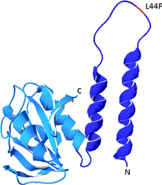
FIGURE 7.
A model of the three-dimensional structure of CdaS based on the known structure of the corresponding YojJ protein of B. cereus (Protein Data Bank code 2FB5) using the SWISS-MODEL homology-modeling server (54). The DAC domain and the N-terminal helices are shown in light blue and dark blue, respectively. The position of the amino acid exchange (L44F) resulting in the hyperactive CdaS protein is highlighted in red.

