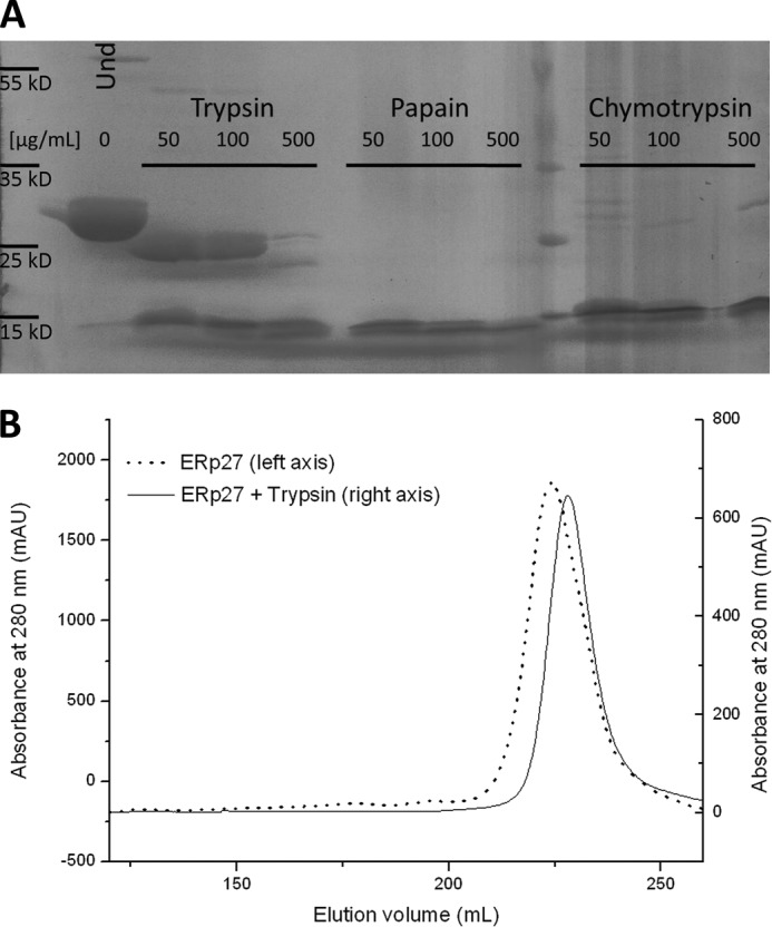
FIGURE 1.

Limited proteolysis of ERp27. A, ERp27 at a final concentration of 500 μm was incubated for 20 min with the indicated proteases at three different concentrations. The left-most lane shows undigested (Und) ERp27 as a control. While both chymotrypsin and papain rapidly degrade the protein, trypsin at lower concentrations only trimmed ∼3 kDa from ERp27. B, comparison of the elution profiles of full-length ERp27 (solid line; left y axis) and the same protein partially digested with 50 μg/ml trypsin (dotted line; right y axis). The fragment generated by proteolytic digestion elutes 5 ml later than the full-length protein and appears as a single species. As only one-third of the full-length protein was subjected to proteolysis, each curve was scaled independently to allow for a better comparison. mAU, milliabsorbance units.
