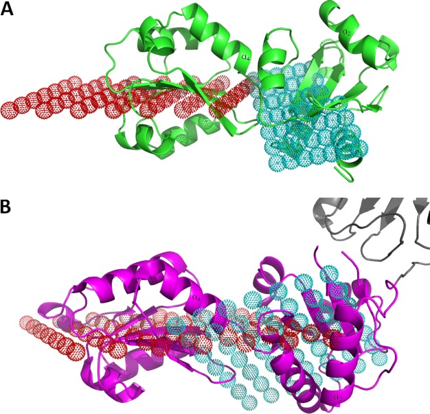
FIGURE 4.
Domain orientation in the PDI family. A, ribbon representation of ERp27 (green) with the reference planes for the N-terminal b domain (red dots) and for the C-terminal b′ domain (cyan dots). Both planes intersect at a shallow angle of 25.7°. B, ribbon model of the bb′ (magenta) fragment of human PDI (Protein Data Bank code 3UEM) with parts of the a′ domain (gray) and the reference planes for the b domain (red dots) and the b′ domain (cyan dots). The angle between the two planes, 55°, is much larger. To allow for a better comparison, one α-helix in each domain has been labeled in A and B.

