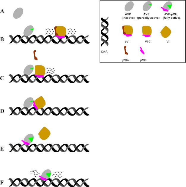
FIGURE 8.
A model, based upon the data in this study, of the activation of AVP by pVI on DNA. A, AVP is inactive, no green mouth. B, AVP bound to DNA is slightly active, green mouth. pVI moves back and forth along the DNA via one-dimensional diffusion. C, pVI slides into AVP. AVP, slightly active, cleaves pVI at its N terminus, releasing the 33-amino acid peptide pVIn. D, a conformational change occurs so that the active site of AVP is at the C terminus of VI-C. E, AVP then cleaves VI-C at its C terminus, releasing the 11-amino acid peptide pVIc that binds to the AVP molecule that cut it out. The fully active AVP-pVIc complex bound to DNA is formed. Protein VI dissociates from the DNA. F, the fully active AVP-pVIc complex bound to DNA slides along DNA via one-dimensional diffusion to locate and process virion proteins also bound to DNA.

