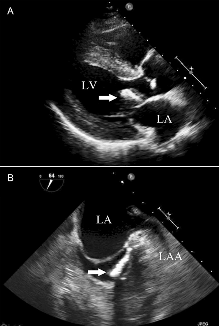
Figure 1:
(A) Transthoracic echocardiogram. The long-axis view shows high-echoic mass (arrow) adhering to the mitral annulus of the anterior leaflet. (B) Transoesophageal echocardiogram. The short-axis view shows a cudgel-shaped, homogenous, high-echoic mass (arrow) that originated from the annulus of the anterior commissure of the mitral valve near the left fibrous trigone. Mitral annulus calcification was also recognized in the same area. LA: left atrium; LV: left ventricle; LAA: left atrial appendage.

