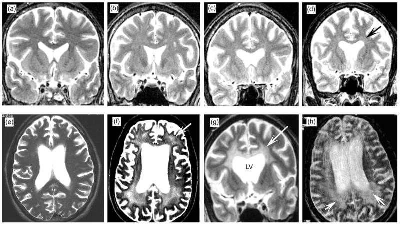
Fig. 1.
White matter alterations by in vivo MRI in patients with HIV-associated leukoencephalopathy. All are T2-weighted images obtained within 12 months of death. The available images were coronal in some but horizontal in others. (a) Normal MRI illustrating the expected characteristics of the cortical ribbon and white matter. (b–d) Cases with mild neuropathology had focal and diffuse white matter hyperintensities (arrow). (e–h) Cases with moderate to severe neuropathology had extensive white matter hyperintensities (arrows). Lesions were most prominent in the white matter surrounding the lateral ventricles (LV), corpus callosum, and optic radiations but were also observed in the frontal pole and parietal white matter.

