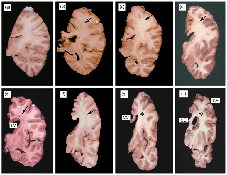
Fig. 2.
Gross findings in the white matter in patients with HIV-associated leukoencephalopathy. All images are coronal sections of the right hemibrain at the level of the hippocampus. The order of the images corresponds to the MRI images in Fig. 1. (a) Normal brain tissue illustrating the white matter and cortical ribbon in a fixed specimen. (b–d) Cases with mild neuropathology had focal white matter lesions in the frontoparietal region (arrows). (e–h) In the most severe cases, the corpus callosum (CC) was diffusely attenuated, the lateral ventricles were dilated, and cavitating lesions were present in the centrum semiovale of the frontoparietal region (arrows). Those with extensive white matter damage also showed cortical atrophy (CA) and reduced basal ganglia volume.

