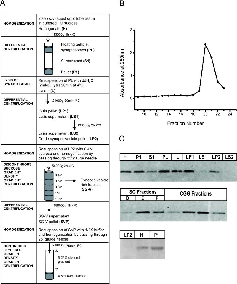
Figure 1.
Synaptic vesicle enrichment by glycerol velocity sedimentation. (A) Scheme summarizing purification of synaptic vesicles from squid optic lobes. (B) Glycerol gradient fractions (250ml) collected from top to bottom and analyzed for their absorbance at 280nm. Major peak indicates synaptic vesicle rich fraction. (C) Electron micrograph of synaptic vesicle rich fraction (D) Western blot of subcellular fractions obtained during the synaptic vesicle purification steps (as described in Materials and Method) with mitochondrial marker; Anti-VDAC (40μg protein/lane) and synaptic marker Anti-SNAP25 (5μg protein/lane) antibodies.

