
Figure 3.
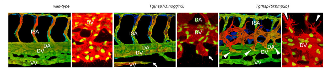
Bmp signaling is necessary and sufficient for sprouting from the axial vein. Blood vessels in wild-type (A, B), Tg(hsp70:noggin3) (C, D) and Tg(hsp70:bmp2b) (E, F) embryos in the Tg(kdrl:GFP) transgenic background (A, C, E). The entire vascular network of 42hpf embryos was analyzed using epiflourescent images; dashed boxes represent the trunk and tail areas analyzed below. Z-stacks from the trunk and tail regions were used to make 3-D color projections (A, C, and E), Filopodia formation of Tg(fli1:nGFP);Tg(kdrl:ras-mCherry) embryos starting at 32hpf (B, D, and F). Arrows in panel c and d show sprouts from the axial vein that fail to make connections in Tg(hsp70:noggin3) embryos. Arrowheads in panel e and f point to ectopic sprouts that branch from the axial vein in Tg(hsp70:bmp2b) embryos. Abbreviations: DA, dorsal aorta; VV, ventral vein; DV, dorsal vein; NC, notocord; NT, neural tube; ISA, intersegmental artery.