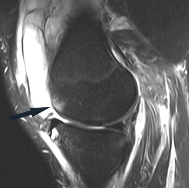
Figure 10.

Sagittal fat-saturated T2-weighted image in a patient with acute grade III posterolateral corner injury shows a relatively small marrow edema pattern in the anterior aspect of the medial femoral condyle (arrow), reflecting a hyperextension-varus injury mechanism. Note the large joint effusion and prominent soft tissue edema.
