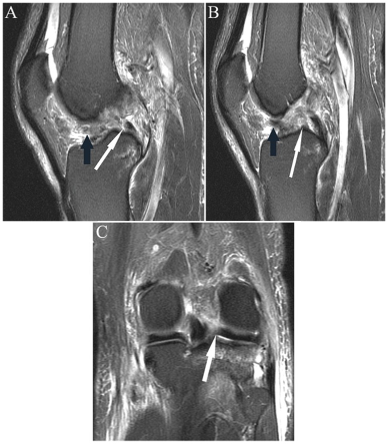
Figure 20.
(A and B) Sagittal and (C) coronal fat-saturated T2-weighted sequences in a patient with acute ACL rupture. Note the focal absence of the anterior portion of the posterior horn root lateral meniscus (white arrow in A) with absence of the expected low signal root in the subsequent more central image (white arrow in B), reflecting root tear/“avulsion.” Figure C shows the tear in the coronal plane (arrow). Note fibers of the ACL displaced anteriorly in the notch (black arrows in A and B). ACL, anterior cruciate ligament.

