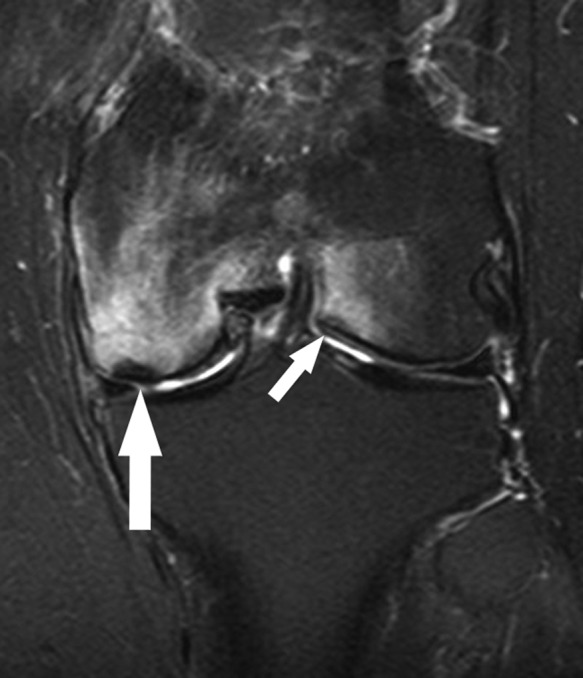
Figure 32.

Six weeks after partial medial meniscectomy, coronal fat-saturated T2-weighted image shows a subchondral fracture of the medial femoral condyle (large arrow) with marked surrounding marrow edema pattern as well as a smaller subchondral fracture of the central aspect of the lateral femoral condyle (small arrow) with less extensive adjacent marrow edema pattern. This case is unusual given subchondral fractures in both compartments; usually, the fracture occurs in the compartment where there has been prior partial meniscectomy or where there is a radial split or complex tear of the posterior horn root.
