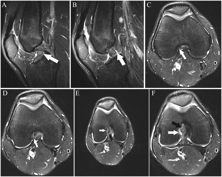
Figure 7.
(A and B) Sagittal and (C-F) axial fat-saturated T2-weighted images show high-grade partial ACL tear. One month prior to the MRI, the patient sustained a knee injury playing soccer. At the time of this MRI, the patient had no pain or subjective instability while running; Lachman, anterior drawer, pivot shift, and KT-1000 measurements were all normal. Three weeks following this MRI, the patient had a twisting injury with positive Lachman and anterior drawer immediately following the injury, consistent with ACL rupture; the ACL was subsequently reconstructed. (A) Abnormal thickening and intermediate signal with undulation of the ACL fibers, most prominent in its midportion (arrow). (B) Abnormal thickening and intermediate signal of the mid fibers with prominent slit-like fluid signal (arrow), reflecting tear. (C) Mild abnormal undulation at the femoral insertion of the anteromedial bundle (arrow). (D) Abnormal intermediate signal and poor delineation of the anteromedial bundle fibers (arrow) is present. (E) Focal fluid signal (white arrow) traversing a portion of the posterolateral bundle at its femoral insertion; there are abnormal disorganized fibers and focal abnormal intermediate signal in the proximal anteromedial bundle (black arrow). (F) Focal disruption of the posterolateral bundle fibers, replaced by fluid signal (white arrow), and abnormal intermediate signal in the anteromedial bundle fibers (black arrow). ACL, anterior cruciate ligament.

