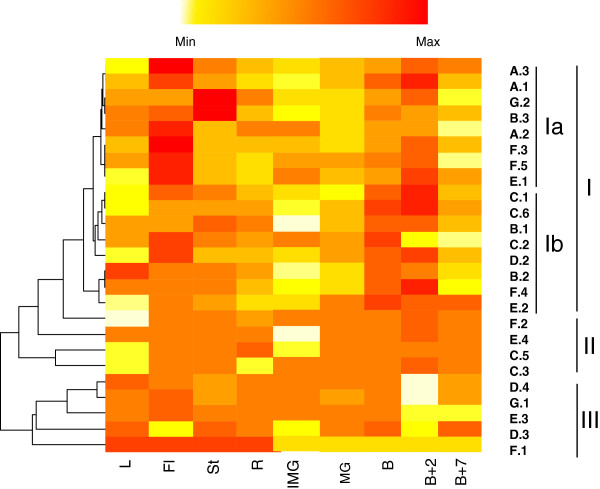
Figure 5.
Heatmap representation of the expression of ERF genes in different tomato tissues. The data obtained by quantitative RT-PCR correspond to the levels of ERF transcripts in total RNA samples extracted from Roots (R), Leaves (L), Stem (St), Flower (Fl), Early Immature Green (IMG), Mature Green (MG), Breaker (B), Breaker + 2 days (B+2), Breaker + 7 days (B+7). The data presented correspond to 3 independent biological repetitions. Red and white colours correspond to high and weak expression of the ERF genes, respectively. Heat map was generated using R software.

