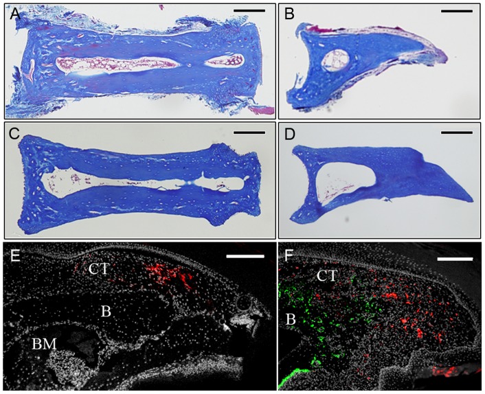
Figure 1. Isolation of connective tissue cells from the P2 and P3 regions.
A, B) Histological section of the P2 (A) and P3 (B) bone after dissection. Loose connective tissue is attached to the surface of the intact bone. C, D) After enzymatic digestion the majority of loose connective tissue was digested off the bone while tissues within the bone marrow are still present. E) DiI labeled cells (red) within the connective tissue (CT) dorsal to the P3 skeletal element (B) are clustered in the amputation stump 2 days post-injection. BM, bone marrow. F) Blastema stage regenerate at 13 DPA showing DiI labeled cells (red) scattered throughout the blastema but not overlapping with Osteocalcin immunohistochemical labeling (green) to identify regenerating osteoblasts. The blastema is contiguous with the connective tissue (CT) and bone (B) of the stump. Scale bars = 200 µm.

