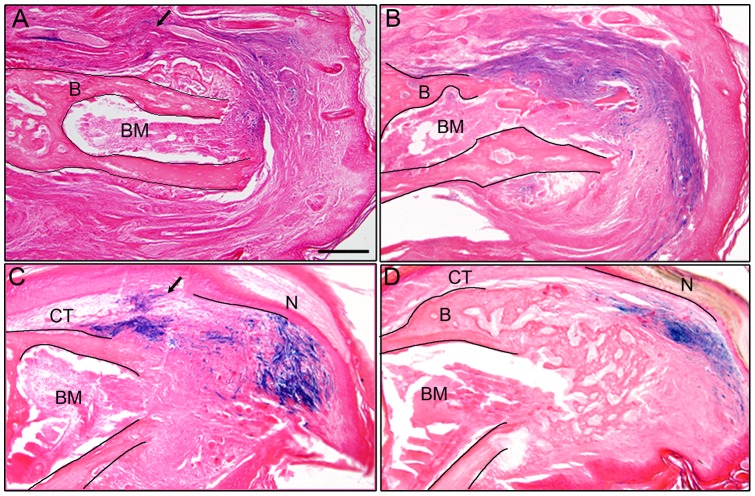
Figure 5. P2 cells participate in a regenerative response.
A) Engraftment of LacZ labeled P3 cells into the dorsal P2 region (arrow) prior to amputation shows labeled cells (blue) at 16 DPA participate with non-labeled dermal cells in the wound healing response. B) Control engraftment of LacZ labeled P2 cells into the dorsal P2 region (arrow) prior to amputation shows labeled cells (blue) at 16 DPA also participate with non-labeled dermal cells in the wound healing response. The amputated P2 bone (B) and bone marrow (BM) are outlined in A and B. C) Engraftment of LacZ labeled P2 cells (arrow) into the dorsal P3 connective tissue (CT) prior to digit amputation shows labeled cells (blue) at 10 DPA participating in blastema formation. D) During the differentiation stage (16 DPA) LacZ positive P2 cells (blue) are found in the regenerating connective tissue (CT) and do not associate with the trabeculae of the regenerating bone. The amputated P3 bone (B), bone marrow (BM) and regenerating nail (N) are outlined in the section to demarcate the stump and the regenerate in C and D. A–D, scale bar = 200 µm.

