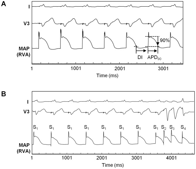
Figure 1. Representative MAP recordings.
Monophasic action potentials (MAPs) recorded at the right ventricular apex (RVA) in a patient with ICM during baseline pacing (A) and during programmed ventricular stimulation (PVS) (B). Basic cycle length (S1–S1) was 500 ms, respectively. (A) Action potential durations (APD) were measured from MAP onset to the 90% repolarization level (APD90). Diastolic interval (DI) span from APD90 of the preceding MAP to the onset of the current MAP. (B) MAP recordings were obtained during PVS using three extrastimuli. In this example, the first two extrastimuli (S2 and S3) were already delivered at the shortest coupling intervals (S1–S2 235 ms, S2–S3 218 ms), while the introduction of the third extrastimulus (S4) was still in progress and the shortest possible S3–S4 interval had not been reached yet.

