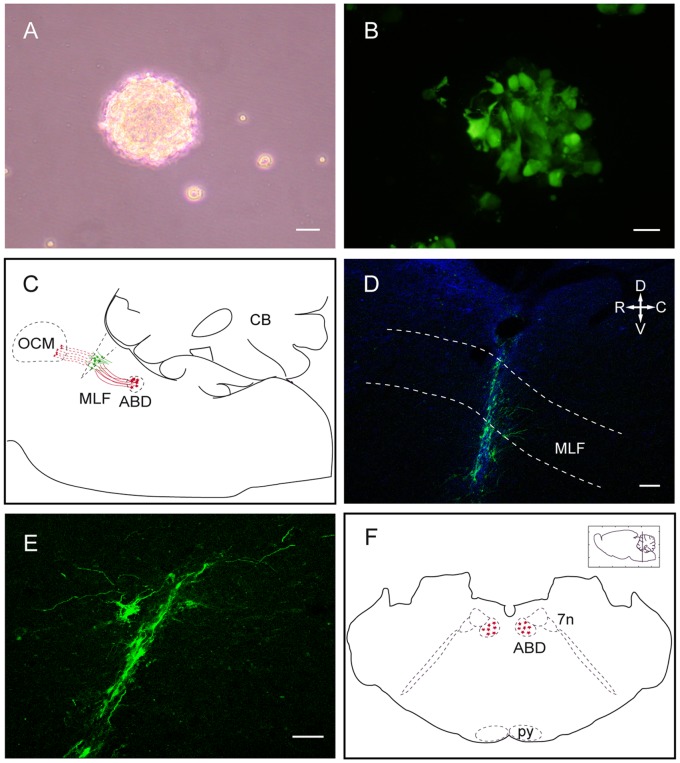
Figure 1. Neural progenitor cell culture, axotomy and cell implant.
A. Floating neurosphere obtained from neural progenitors of the postnatal rat subventricular zone (SVZ). Scale bar: 25 µm. B. GFP-expressing cells (in green) in a SVZ-derived neurosphere. Scale bar: 25 µm. C. Schematic drawing of a rat parasagittal brainstem section showing the location of the medial longitudinal fascicle (MLF) transection and cell implant. Abducens internuclear neurons are represented in red and implanted neural progenitor cells in green. Axons of abducens internuclear neurons (red lines) course through the MLF towards the contralateral oculomotor nucleus. The distal stump of disrupted axons are represented in red dashed lines. D. Confocal microscopy image of a parasagittal brainstem section showing the implanted cells labeled with GFP (in green) at the site of axotomy. Dashed lines indicate the approximate dorso-ventral limits of the MLF. Scale bar: 100 µm. E. Higher magnification image of implanted GFP-labeled cells. Dorso-ventral and rostro-caudal orientation as in D. Scale bar: 50 µm. F. Schematic representation of a rat coronal section through the pons showing the abducens nucleus location. Abducens internuclear neuron somata are represented in red. Abbreviations: ABD: abducens nucleus; C: caudal; CB: cerebellum; D: dorsal; GFP: green fluorescent protein; MLF: medial longitudinal fascicle; OCM: oculomotor nucleus; py: pyramidal tract; R: rostral; V: ventral; 7n: facial nerve.

