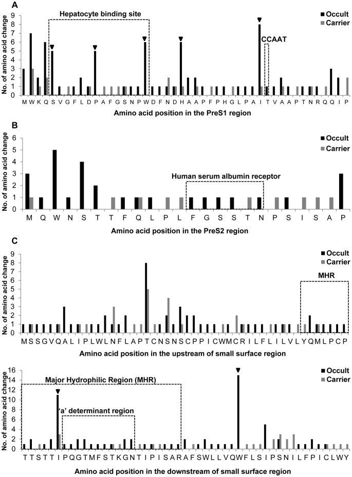
Figure 3. Location of mutated codons in the preS1 (A), preS2 (B) and S region (C) and comparison of the mutation frequencies between occult subjects and carriers.
The inverted triangle (▾) indicates codons for which substitutions were significantly or tended to be significantly prevalent in the occult HBV subjects as compared to the carriers.

