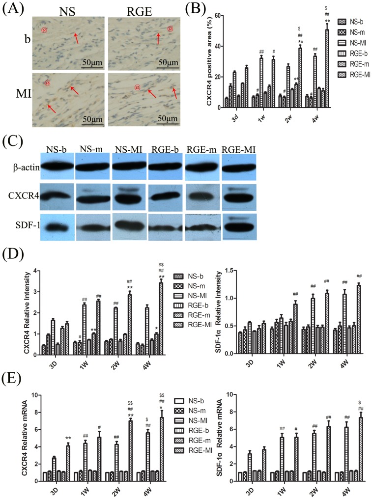
Figure 6. The expression of the stromal-derived factor-1α (SDF-1α) and its receptor 4 (CXCR4) in tissue.
(A) Immunohistochemistry of CXCR4 in NS-b, NS-MI, RGE-b and RGE-MI groups at week 4. The vessels are indicated by red @, and positive colorations are indicated by red arrows. (B) Western blot analysis of protein expression of CXCR4 and SDF-1α. β-actin was an internal reference protein for Western blot. (C) Quantitative of CXCR4 immunoreactivity (percentage of positive area) in each group. (D) Quantitative Western blot analysis of CXCR4 and SDF-1α in myocardial tissue. (E) Quantitative PCR analysis of CXCR4 and SDF-1α in myocardial tissue. Data are mean ± SD. *P<0.05, **P<0.01 vs. NS group at the same time, #P<0.05, ##P<0.01 vs. the same group at day 3, $P<0.05, $$P<0.01 vs. the same group at week 1.

