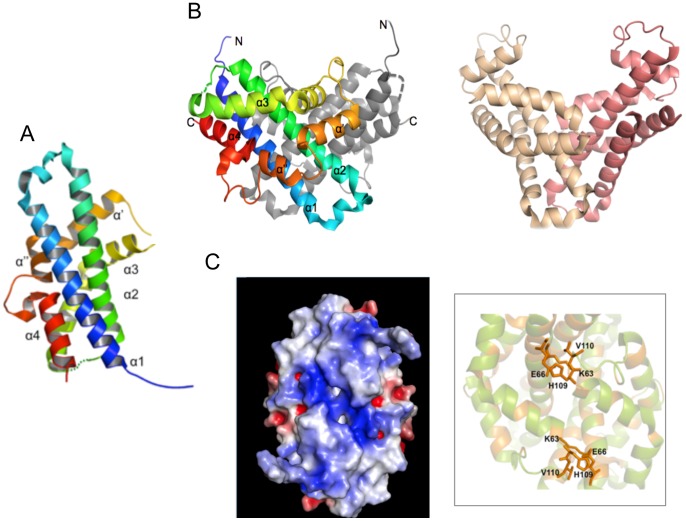
Figure 3. The Vpc_cass2 dimer (PDB 3JRT).
A. Ribbon of monomeric unit with colour spectrum (blue to red) across six helical components (α1, α2, α3, α′, α″, α4), named as indicated. A loop of weak density connecting helices 2 and 3 is represented by dotted line. B. Contrasting shapes of dimers of Vpc_cass2 (left panel) and structural relative HI0074 from Haemophilus influenza [55] (right panel). C. View (left panel) across Vpc_cass2 dimer interface shown as electrostatic surface (in blue to red from +5 to −5 kbT/e [57]), highlighting the basic cluster unique to this protein. Right panel provides segmental view of dimer ribbon (green) and side chains of putative active site residues of Vpc_cass2 straddling the dimer interface. Residues conserved in Vpc_cass2 and its sequence homologs from Shewanella and Moritella sp are coloured orange.

