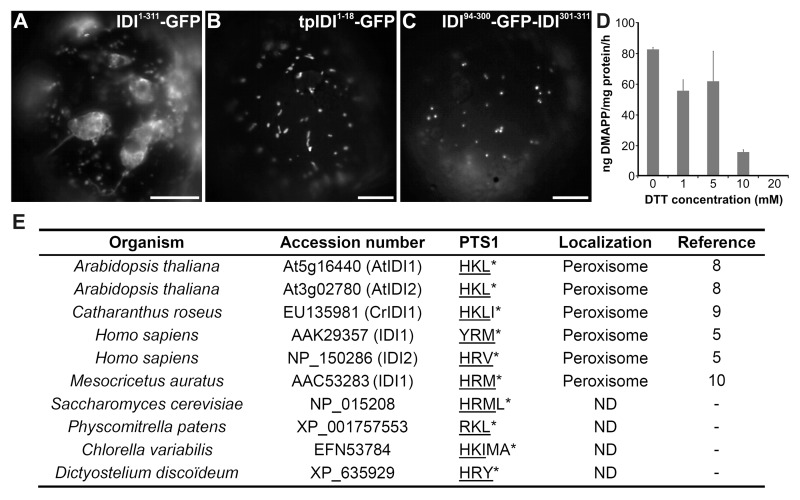
Figure 1. Subcellular localization of IDIs. (A) Dual plastid and mitochondrial targeting of the long isoform of CrIDI1 (residues 1 to 311) fused to GFP (IDI1–311-GFP). (B) Mitochondrial targeting of the first 18 residues of the CrIDI1 transit peptide fused to GFP (tpIDI1-18-GFP). (C) Peroxisomal targeting of the CrIDI1 short isoform bearing an internally fused GFP (IDI94–300-GFP-IDI301–311). (D) Effect of DTT on CrIDI1 activity as judged by incubation of IPP with CrIDI1 at different DTT concentrations. IPP isomerized to DMAPP was detected as isoprene gas following its acidification with phosphoric acid as previously described.15 Error bars signify standard deviation of two replicates. (E) Sequences of PTS1 and putative PTS1 in IDIs from different organisms. The tripeptide of PTS1 is underlined. The asterisk indicates the end of the polypeptide chain. Peroxisomal localization is indicated when experimentally validated. Accession numbers are from GenPept and from Arabidopsis Genome Initiative (for AtIDI1 and AtIDI2). ND, not determined; Bars, 10 µm.

An official website of the United States government
Here's how you know
Official websites use .gov
A
.gov website belongs to an official
government organization in the United States.
Secure .gov websites use HTTPS
A lock (
) or https:// means you've safely
connected to the .gov website. Share sensitive
information only on official, secure websites.
