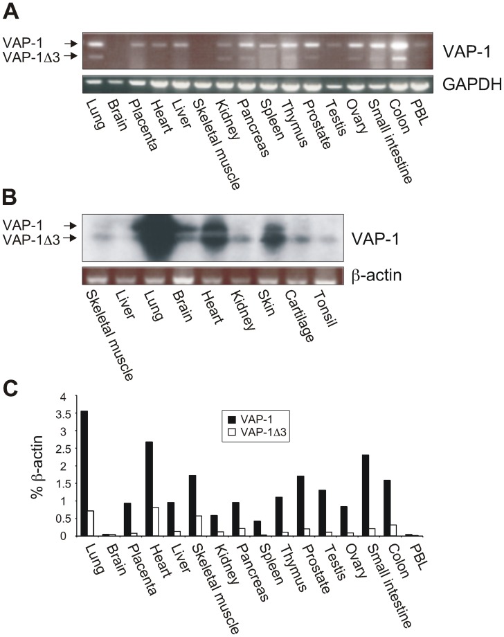
Figure 2. Expression of VAP-1Δ3 mRNA in adult and fetal human tissues.
(A) Commercial first strand cDNA panels were used to determine the mRNA expression of VAP-1Δ3 in respect to the expression of the full-length VAP-1 in adult human tissues by PCR. GAPDH expression was used as an endogenous control. (B) RT-PCR analysis of nine fetal human tissues was performed using VAP-1 and β-actin specific primers. The identity of the resulting amplicons was verified with Southern blotting using a VAP-1-specific probe. The two alternatively spliced mRNA species are marked with arrows. (C) qPCR analysis was performed with transcript specific primers and probes using the commercial first strand cDNA panels as templates. The expression levels of VAP-1 and VAP-1Δ3 are presented as percentages of β-actin mRNA expression in the same sample.

