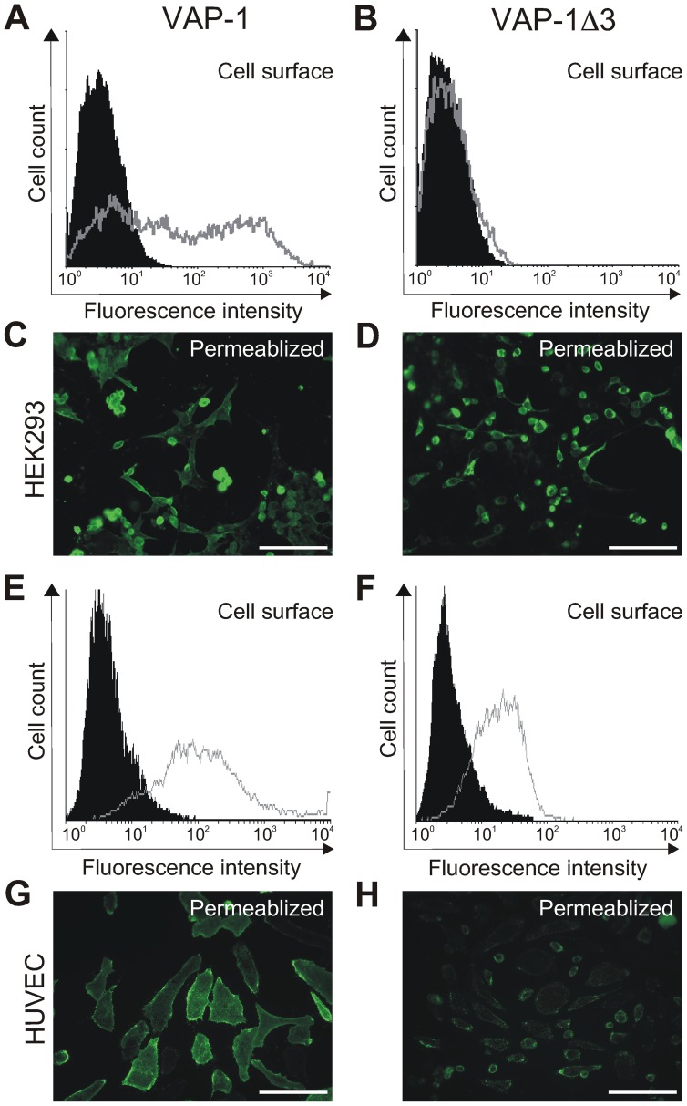
Figure 3. Expression of VAP-1Δ3 in transiently transfected cell lines.
Flow cytometry of HEK293 cells transfected either with the full-length VAP-1- or with VAP-1Δ3 -cDNAs in pcDNA3.1 (A–B). The gray histograms: staining with the anti-VAP-1 polyclonal antibody; the black histograms: staining with a negative control antibody. The expression was also examined by fluorescence microscopy of acetone-permeabilized coverslip-plated HEK293 cells transfected with the corresponding constructs (C–D). In E–F, flow cytometry of HUVECs infected with pAdCMV-constructs of VAP-1- and VAP-1Δ3. The gray histograms: staining with the anti-VAP-1 polyclonal antibody; the black histograms: staining with a negative control antibody. The expression was also examined by fluorescence microscopy of acetone-permeabilized coverslip-plated HUVECs infected with the corresponding constructs (G–H). Scale bar 100 µm.

