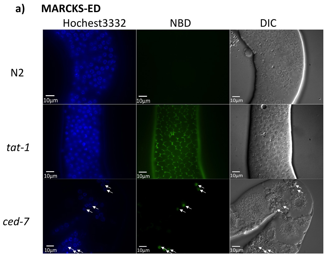
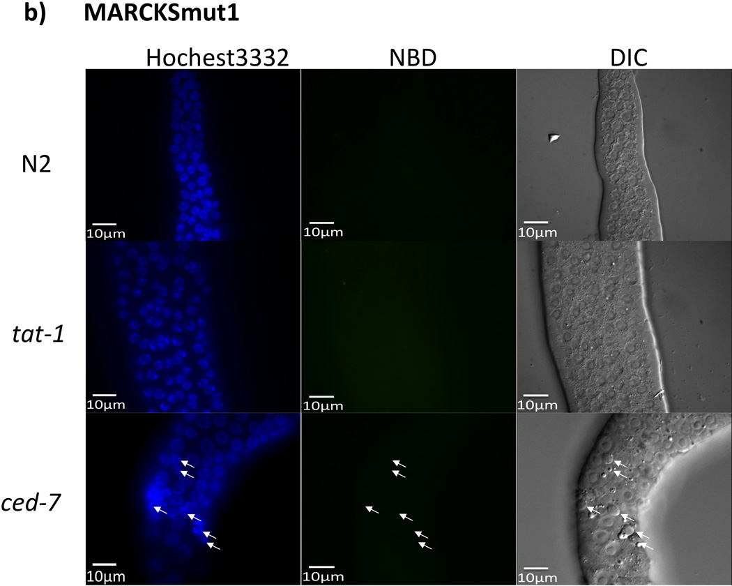
Figure 4.


In vivo C. elegans fluorescence assay. The exposed gonads of a wild type N2 hermaphrodite animal (top row), a tat-1(tm3117) mutant animal (middle row), and a ced-7(n2094) mutant animal (bottom row), were stained with NDB-labeled a) MARCKS-ED or b) MARCKSmut1. Images of Hoechst 33342 staining (cell nucleus), MARCKS peptide staining, and differential interference contrast (DIC) microscopy (22) are shown. Arrowheads indicate apoptotic cell corpses. Scale bar = 10 µm.
