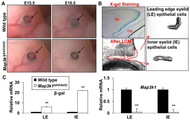
Figure 1. MAP3K1 expression and function in developing eyelids.
(A) Photographs show the developing eyes of wild type and Map3k1ΔKD/ΔKD fetuses at E15.5 and E16.5. Arrows point at developing eyelids, showing that the eyelids are fully open in E15.5 fetuses of both genotypes, while they are closed in E16.5 wild type, but not knockout fetuses. (B) X-gal stained Map3k1 heterozygous fetuses (Map3k1+/ΔKD) at E15.5 were sectioned and photographed (upper left). Pictures were taken before (upper left) and after (lower left) LCM, and the captured leading edge eyelid (LE) (upper right) and inner eyelid (IE) (lower right) epithelium. (C) Total RNA from LE and IE epithelium were subjected to qRT-PCR using primers for the kinase domain of Map3k1 (Map3k1) and β-gal as indicated. Relative expression was calculated based on that of Gapdh in each sample, and compared to the expression in wild type cells, set as 1. The results are shown as mean ± SD from at least 3 samples of each genotype and triplicate PCR of each sample. Statistic analyses were done by Student t-test, **p < 0.01 is considered significant. LE, leading edge eyelid, IE, inner eyelid, DE, dermis, CO, cornea.

