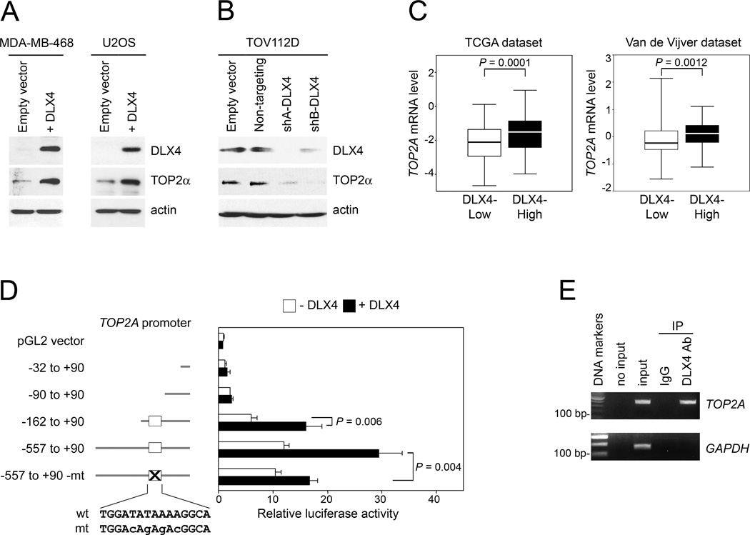
Fig. 1. DLX4 induces TOP2A promoter activity and TOP2α levels.
[A,B] Western blot of DLX4 and TOP2α levels in [A] vector-control and DLX4-overexpressing MDA-MB-468 and U2OS cell lines, and [B] TOV112D cells transfected with empty vector, non-targeting shRNA and shRNAs targeting two different sites of DLX4 (shA-DLX4, shB-DLX4). [C] Breast cancer cases from the TCGA Project (n=537) and study of Van de Vijver et al (32) (n=295) were stratified according to DLX4 expression in tumors, where DLX4 transcript levels in each dataset were defined as High (> upper quartile) and Low (< lower quartile). Significance of differences in TOP2A transcript levels (log2 scale) between upper and lower quartile sub-groups was evaluated by Mann-Whitney U-test. [D] U2OS cells that lacked DLX4 (white bar) or expressed DLX4 (black bar) were transfected with pGL2 luciferase reporter plasmids containing the indicated regions of the TOP2A promoter. Wild-type (capitals) and mutated (small case) sequences of the DLX4-binding site (white box) are indicated. Shown are average relative luciferase activities of 3 independent experiments. [E] Detection of binding of endogenous DLX4 to the TOP2A promoter in TOV112D cells by chromatin IP. Input corresponds to 1% of the chromatin solution before IP.

