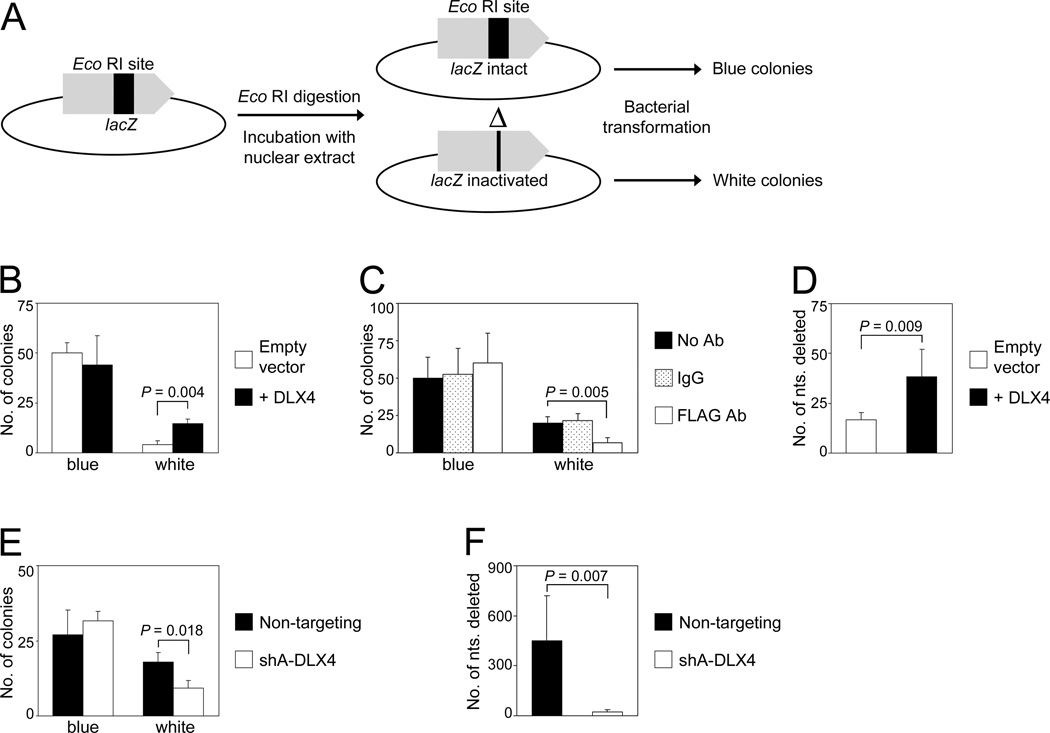
Fig. 3. DLX4 stimulates erroneous end-joining repair of DSBs.
[A] Schematic diagram of the end-joining assay. Repair of the Eco RI-cut lacZ gene of pUC19 DNA incubated in nuclear extracts was assayed by formation of blue colonies (correct repair) and white colonies (misrepair) following transformation of E. coli and plating on plates containing X-gal and IPTG. [B] Colony formation in end-joining assays using nuclear extracts of vector-control U2OS cells and U2OS cells that expressed FLAG-tagged DLX4 (+DLX4). [C] Colony formation in end-joining assays using nuclear extracts of U2OS cells that expressed FLAG-tagged DLX4, where extracts were depleted using FLAG Ab, IgG or left untreated. [D] Numbers of nucleotides surrounding the Eco RI-cut DSB that were deleted in pUC19 DNA isolated from individual white colonies of end-joining assays using U2OS extracts. [E] Colony formation in end-joining assays using nuclear extracts of TOV112D cells transfected with non-targeting and DLX4 shRNAs. [F] Numbers of deleted nucleotides in pUC19 DNA isolated from white colonies of end-joining assays using TOV112D extracts. Shown are average results of 3 independent experiments.

