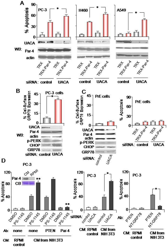
Figure 4. UACA knock-down induces apoptosis by Par-4.

(A,B) UACA knock down affects cancer cells. The indicated cell lines were transfected with UACA siRNA or control siRNA duplexes. As indicated in Figure S4, transfection with two different siRNA duplexes and one shRNA was used to confirm the findings. (B) Up-regulation of GRP78 at the cell surface was determined by FACS analysis of unfixed cells. (A) The cells were treated with TRX or TRX-Par-4 (100 nM) for 24 h, and subjected to ICC for active caspase 3 and scored for apoptosis. Knock-down of UACA was confirmed by Western blot (WB) analysis.
(C) UACA knock-down does not affect normal cells. PrE cells were transfected with siRNA duplexes, and up-regulation of GRP78 at the cell surface was determined by FACS analysis of unfixed cells (left panel), knock-down of UACA was confirmed by Western blot (WB) analysis (left panel), or the cells were treated with TRX or TRX-Par-4 (100 nM) for 24 h, and scored for apoptosis (right panel).
(D) Effects of Par-4 protein secreted by mammalian cells. PC-3 cells were treated with CM from NIH 3T3 fibroblasts or with RPMI control medium, in the presence of PS-1145 or DMSO, and no antibody, PTEN antibody or Par-4 antibody (left panel), or GRP78 N-terminal antibody or control antibody (right panel). Moreover, the cells were transfected with UACA siRNA or control siRNA duplexes, then treated with CM from NIH 3T3 fibroblasts or with RPMI control medium for 24 h, and scored for apoptosis (middle panel). Par-4 in the CM was confirmed by Western blot analysis, using albumin in the Coomassie blue (CB) stained gel to normalize loading (left panel, inset). Each treatment used 5 nM secreted Par-4 protein, as judged by quantitative Western blot analysis (not shown).
Asterisk (*) indicates statistically significant (P < 0.001) difference by the Student t test; and (**) indicates that the effect is significant (P < 0.001) based on two-way ANOVA with data normality and equality of variance assumptions.
