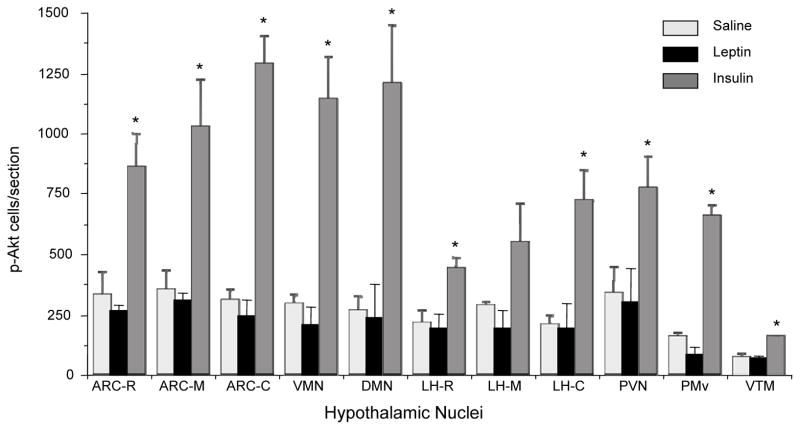
Fig. 6.
Changes in the number of p-Akt (Ser473) positive cells in various hypothalamic sites of saline, leptin or insulin-treated rats. One of six series from each animal was analyzed. ARC, arcuate nucleus; VMN, ventromedial nucleus; DMN, dorsomedial nucleus; LH, lateral hypothalamus; PVN, paraventricular nucleus; PMv, ventral premammillary nucleus; VTM, ventral tuberomammillary nucleus. R, M and C for respective nuclei represent rostral, middle and caudal part, respectively. Values represent the mean ± SEM for 3 animals per group.*P < 0.05 vs. saline group.

