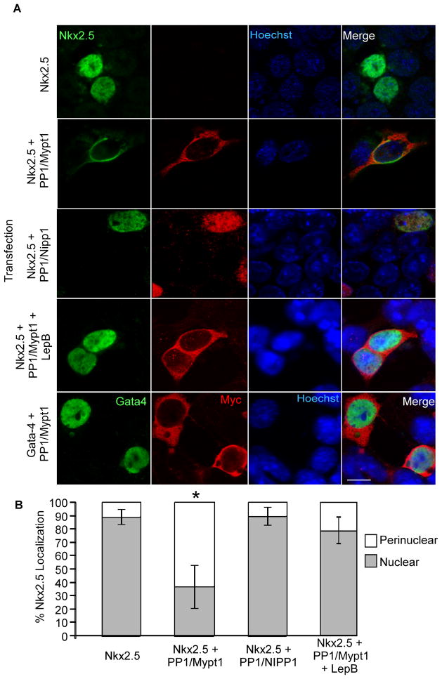
Figure 2. Coexpression with MP results in exclusion of Nkx2.5 from the nucleus.
P19 cells were transiently transfected with the plasmids indicated and fixed one day later for analysis of subcellular localization. A) Fixed cells were labelled with antibodies specific to Nkx2.5 and myc-tag as well as with Hoechst dye to detect nuclei. Scale bar represents 10μm. B) Subcellular localization of Nkx2.5 was quantified by imaging 10 fields in three independent experiments. An average of 142 cells were quantified per treatment (n=3, *p<0.05).

