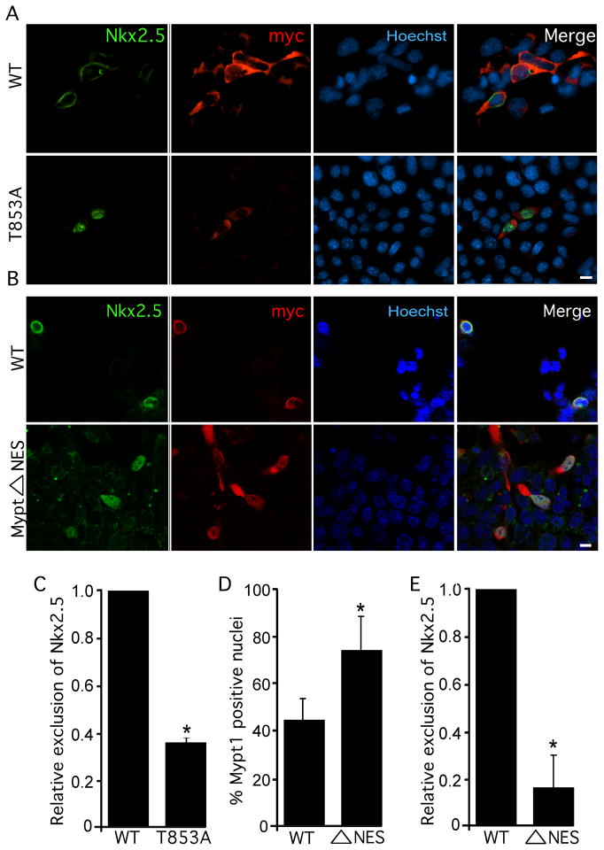
Figure 4. Thr853 and a nuclear export signal in Mypt1 are required for nuclear exclusion of Nkx2.5.
P19 cells were transiently transfected with Flag-Nkx2.5, HA-PP1β and myc-Mypt1, myc-Mypt1Δ853A (A, C) or myc-Mypt1 NES (B, D, E). A &B) Cultures were labelled by immunofluorescence with antibodies specific to Nkx2.5 and myc as well as with Hoechst dye to detect nuclei. Scale bar represents 10μm. C & E) The subcellular localization of Nkx2.5 was quantified in wild-type, Mypt1T853A, and myc-Mypt1ΔNES expressing cultures (n=3, *p<0.05). D) The subcellular localization of Mypt1 and Mypt1ΔNES was quantified (n=3, *p<0.05).

