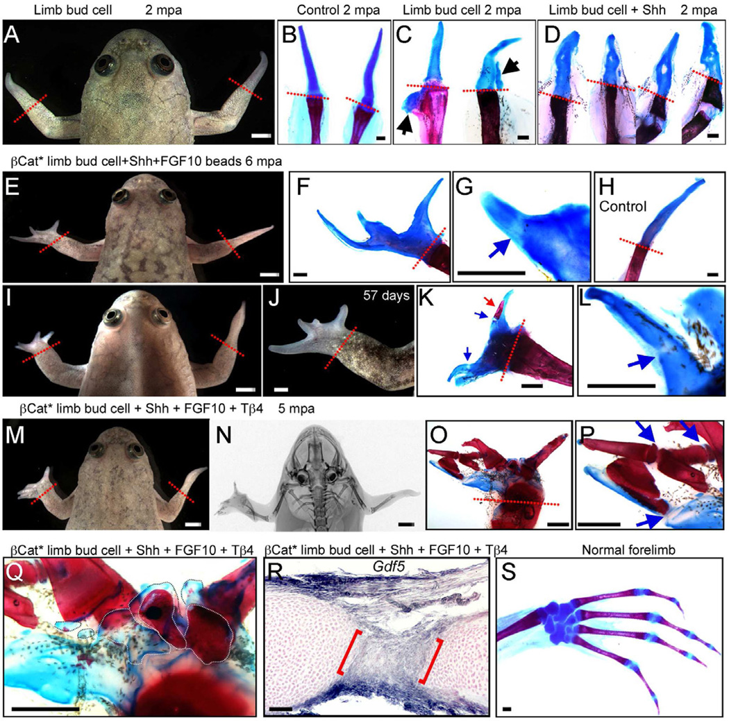
Fig. 3. Xenopus frog forelimb regeneration after cell transplantation.
Left limbs are transplanted and right limbs are controls with amputation only.
(A –C) Limb regeneration in a frog with a GFP limb bud cell patch, 2 months post amputation (mpa). Skeletal staining shows that regenerates are still simple spikes (A, C) similar to controls (B), although 2 cases gave slight extra cartilage (arrows in C).
(D) Skeletal preparations of 4 forelimbs treated with limb bud cell patches and Shh beads, showing slightly disturbed cartilages, 2 mpa.
(E–L) Frogs treated with a βcat* limb bud cell patch and Shh + FGF10 beads (BSF), 6 mpa. Multidigit regenerates are formed (E, F, I, J) and some proximodistal segmentation of cartilage is evident (blue arrows in G, L). One digit also has a small patch of calcium deposition (red arrow in K). All panels except (H) are for BSF treatment. (H) shows skeletal preparation of a typical spike as control, 6 mpa.
(M–Q) A frog treated with a βcat* limb bud cell patch, Shh + FGF10+ thymosin β4 (BSFT), 5 mpa. Multiple digits have regenerated (M) and X-ray shows the presence of extensive ossification in the regenerate, as confirmed by skeletal staining (O, P). Some proximodistal segmentation is also clear (blue arrows in P). (Q) Regeneration of intermediate skeletal elements in frog shown in (M). White dotted lines indicate individual metacarpal-like structures.
(R) Detection of Gdf5 by in situ hybridization in regenerate from frog treated with BSFT. Cross section of a digit (2 mpa) shows joint-like structure, with Gdf5 expression (between red brackets).
(S) A skeletal preparation of a normal forelimb, for comparison.
Red dotted lines indicate amputation levels. Scale bars in (A, E, I, J, M, N), 500 µm; Scale bars in (B-D, F-H, K, L, O–S), 100 µm.
See also Fig. S3 and S4.

