Fig. 1.
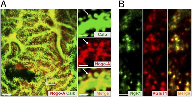
Distribution of Nogo-A and NgR1 in the cerebellum. (A and B) Nogo-A and its receptor NgR1 are complementarily expressed at synaptic sites in the cerebellum of P28 WT/BL6 mice. (A) Double immunolabeling with anticalbindin (green) and anti–Nogo-A (red) antibodies revealed the presence of Nogo-A in dendrites and spines (arrows) of PCs. Higher power micrographs correspond to the boxed region. (Scale bars, 5 µm; 2 µm in Insets.) (B) Colabeling with antibodies against NgR1 (green) and VGluT1 (red) demonstrates the strong expression of NgR1 in the presynaptic, VGluT1+ PF terminals. (Scale bar, 2 µm.)

