Fig. 3.
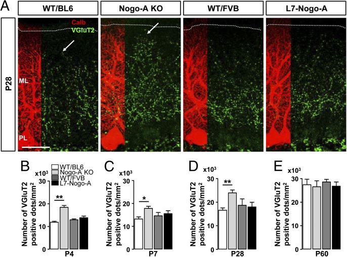
Genetic deletion of Nogo-A affects the developmental rearrangement of CF terminals on PC dendrites. (A) Confocal images of VGluT2+ CF terminals (green) on P28 PCs stained for calbindin (red). Note the increased territory of CF synapses in Nogo-A KO mice at P28 (arrows). Dotted lines indicate the pial surface. ML, molecular layer; PL, PC layer. (Scale bar, 50 µm.) An increase in the density of VGluT2+ CF varicosities was found in Nogo-A KO mice at P4 (B), P7 (C), and P28 (A and D). This effect of Nogo-A KO on the CF innervation pattern was no longer visible at P60 (E). Overexpression of Nogo-A had no effect on either the density or the position of CF terminals (A–E). Values represent means ± SEM of 103–116 cells (six mice) per genotype; *P < 0.05, **P < 0.01, Student t test.

