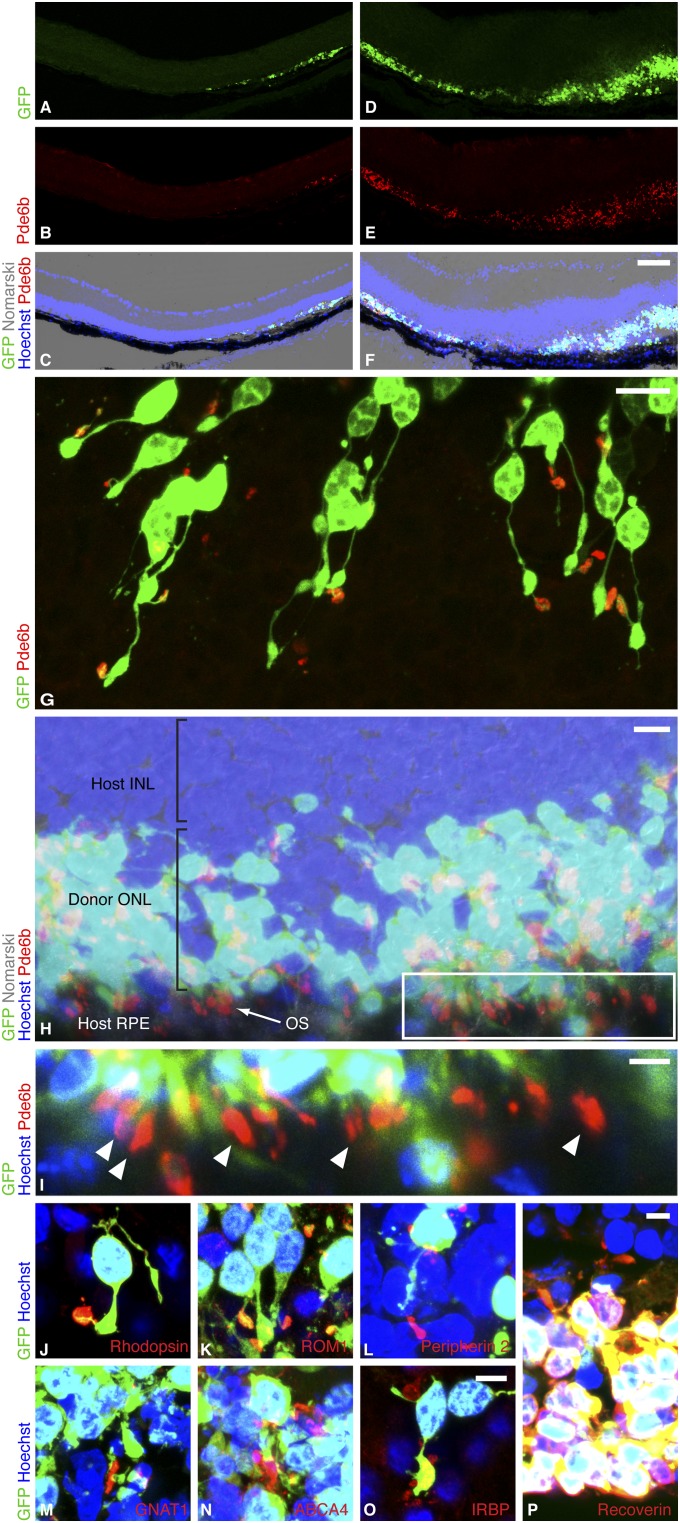
Fig. 1.
Rod precursors transplanted into the degenerate host regenerate a new photoreceptor layer. (A–F) Two regions of rd1 host retina after subretinal transplantation of P3 Tg(Nrl-L-EGFP) rod precursors. By 2 wk, the retina had reattached and a new donor-derived ONL, marked by GFP, was formed. A–C show an area where one to three ONL rows were formed, and D–F show up to 10 rows. An internal nontransplant control region lacking an ONL is shown in the left half of A–C. (Scale bar, 75 µm.) (G) As a sign of maturation, donor-derived GFP-positive rods formed slender processes extending from the cell body and expressed phosphodiesterase β6 (Pde6b) sequestered in discrete OS. (Scale bar, 10 µm.) (H) In many regions, Pde6b-positive OS were appropriately polarized with respect to host RPE, which is the optimal configuration for rod OS maintenance. (Scale bar, 10 µm.) Boxed region of H enlarged in I shows OS (arrowheads) aligned in rows against host RPE. (Scale bar, 5 µm.) (J–N) As further evidence of maturation, rhodopsin, rod outer segment membrane protein 1 (ROM1), peripherin 2, guanine nucleotide-binding protein G(t) subunit α1 (GNAT1), and ATP-binding cassette subfamily A member 4 (ABCA4) were localized in outer segments. (O) Donor cells expressed interphotoreceptor retinoid-binding protein (IRBP), a secreted protein critical for ONL maintenance, and (P) were positive for the photoreceptor marker recoverin. (Scale bar, 5 µm for J–P.)

