Fig. 1.
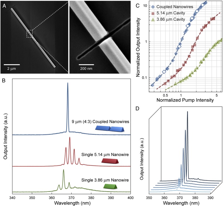
Single-frequency lasing in 9-μm (4:3) cleaved-coupled nanowires. (A) SEM images showing the axially coupled GaN nanowires on a Si substrate with 250-nm–thick thermal oxide. The two-component nanowires are ∼100 nm in radius and coupled via an intercavity gap ∼40 nm wide. (B) Single-wavelength lasing is observed from the coupled cavity (blue line), and each component nanowire lases at multiple wavelengths when they are separated. (C) Clear lasing transitions can be seen from the power-dependent measurements. The pump intensity and the output intensity are both normalized to the values at the threshold of the cleaved-coupled nanowires. The dashed lines were fitted using a rate equation analysis (SI Appendix, section S11). (D) Output spectra of the cleaved-coupled nanowires show clean single-wavelength lasing at different excitation intensities corresponding to the open diamonds in C.

