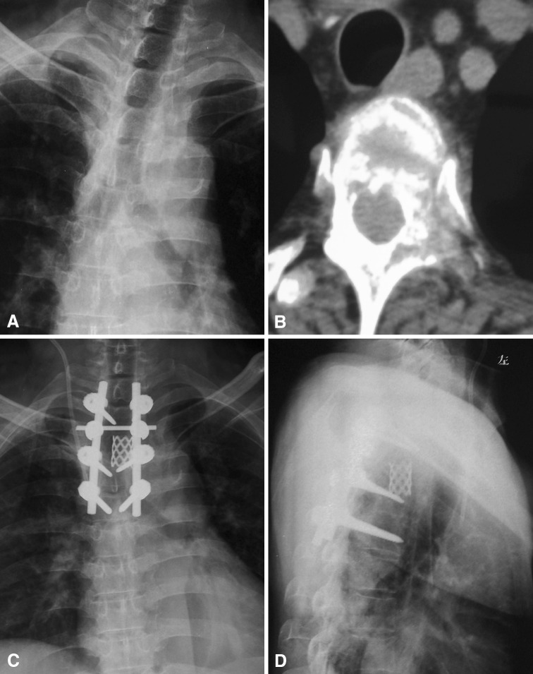
Fig. 4A–D.
A 65-year-old man previously diagnosed with prostate cancer had severe chest and back pain and weakness in both lower extremities. His radiograph and CT results revealed the T3 vertebra had been destroyed by an invasive mass and the spinal cord was compressed. Posterior total en bloc resection of the T3 tumor, bone graft fusion with titanium mesh, and pedicle screw fixation were performed. His pain was relieved and neurologic function was improved postoperatively. The patient was still alive at 41 months after the surgery with no recurrence. (A) A preoperative radiograph shows a destroyed T3 vertebra and vertebral pedicles, and (B) his transverse CT scan shows an invasive mass that has destroyed the vertebra and invaded the paravertebral tissue and spinal canal, resulting in spinal cord compression. (C) AP and (D) lateral view postoperative radiographs show stable fixation.

