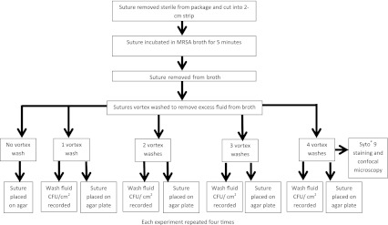
Fig. 1.

A diagram of the experimental design is shown. Each suture was placed in MRSA broth for 5 minutes. The sutures were removed and underwent a vortex wash in normal saline for 15 seconds. The wash solution then was examined and CFU/mL recorded. The suture was placed on an agar plate and cultured overnight. Sutures from the four vortex wash groups underwent Syto® 9 DNA staining and were examined under confocal microscopy to determine adherence patterns (CFU = colony forming units).
