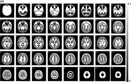
Fig. 3.


a X-ray CT image of the developed phantom, which does not contain water or bone-equivalent liquid inside the phantom. Note that the images are converted to attenuation μ value with a peak value of 0.10 (cm−1). The images are aligned to the digital design shown in Fig. 2. No remaining materials or irregular surface are seen inside the grey matter or bone compartments of the phantom. b X-ray CT images of a phantom that contains water in the grey matter region, and a K2HPO4 solution in the bone compartment. Note that the images are converted to attenuation μ value with a peak value of 0.20 (cm−1). The images are aligned to the digital design of Fig. 2. There are no air bubbles remaining in the phantom
