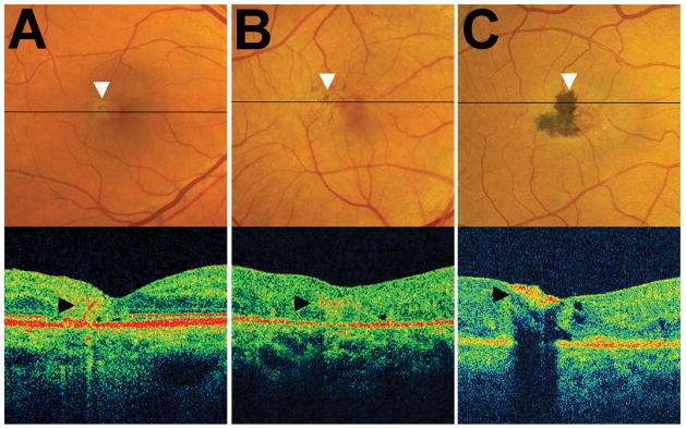
Figure 4. Cross-sectional localization of pigment clumping within the neuroretina using optical coherence tomography (OCT).

Horizontal OCT B-scans were obtained in the retinal plane traversing an area of pigment clumping (indicated by a black horizontal line) in individual study eyes. (A) Example of a study eye with a small isolated island of pigment in the temporal perifoveal region. OCT demonstrates a band of hyperreflectivity (arrowhead) that originated from the RPE layer and extended into the inner layers of the retina. (B) Example of a study eye with multiple small islands of pigment clumping which on OCT are observed as a horizontal area of reflectivity within the retina at the level of the outer plexiform layer or inner nuclear layer (arrowhead). (C) Example of a study eye with a large contiguous area of pigment clumping which appears on OCT as a hyperreflective plaque on the inner aspect of the retina (arrowhead).
