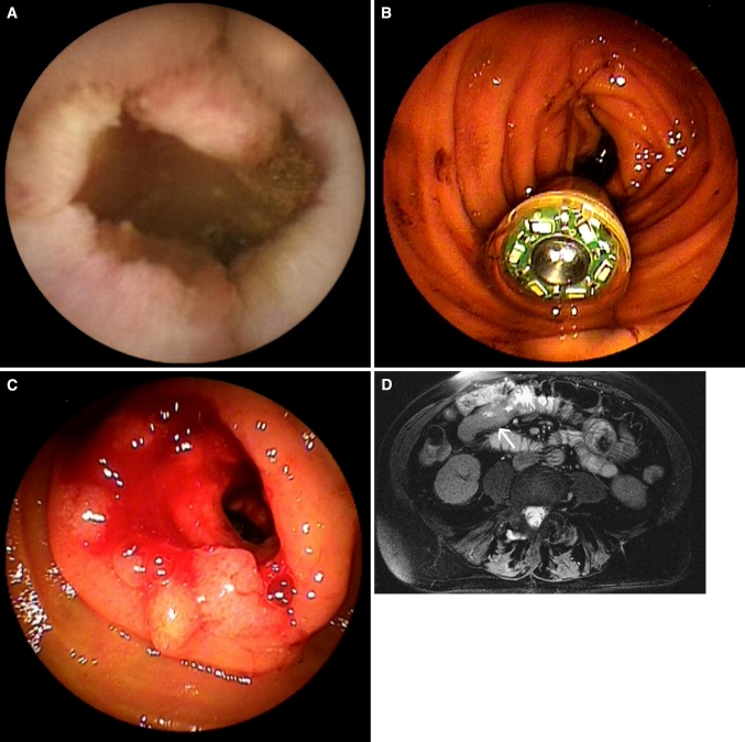
Fig. 1.
82-Year-old female with iron-deficiency and negative conventional bidirectional endoscopy. A Capsule image showing an irregular stenotic mass lesion. B Emergency double-balloon endoscopy was performed because of obstructive symptoms 6 h after ingestion of the capsule and showed the capsule in the proximal small intestine. C After endoscopic removal of the capsule, a stenotic mass lesion became visible. Biopsy specimens revealed the lesion to be carcinoma. D Transverse True FISP MR enteroclysis image showing wall thickening and obstruction of the proximal jejunal lumen (arrow).

