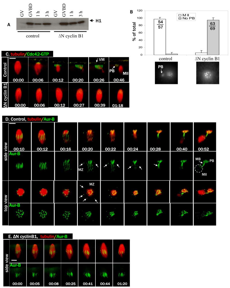
Figure 1. Cyclin B degradation is required for anaphase initiation and for Cdc42 activation.
A. Control oocytes and oocytes injected with ΔN cyclin B1 mRNA were incubated with progesterone and withdrawn at GVBD, 1h or 3h after GVBD, for MPF (histone H1 kinase) assays. GV oocytes were not treated with progesterone. Note the transient (1h) inactivation of MPF in control but not ΔN cyclin B1 oocytes.
B. Control oocytes and oocytes injected with ΔN cyclin B1 mRNA were treated with progesterone and fixed 3–4 h after GVBD for chromosome analyses. Oocytes were scored as MII (metaphase II chromosome array and a polar body) or no polar body (PB). Shown are means (% of total oocytes examined, with SEM) of three independent experiments. The total numbers of oocytes in the three experiments are also included in the graph. Shown below are typical chromosome images of the two groups.
C. A series from 4D movie of a control oocyte (upper row) and an oocyte injected with ΔN cyclin B1 mRNA (lower row). These oocytes were also injected with rhodamine-tubulin (red) and eGFP-wGBD (active Cdc42, green). Note the complete lack of Cdc42 activation, and lack of a polar body (PB), in oocytes injected with ΔN cyclin B1 mRNA. Scale bar is 20 μm in all images of this paper. Time (hr:min:sec) zero corresponds to the beginning of the time lapse experiments, typically 100–120 min after GVBD. In some oocytes, the vitelline membrane (VM) is visible with the fluorescence probes.
D. A series from 4D movie of a control oocyte depicting dynamic localization of endogenous Aurora B (Alexa 488 anti-Aurora B, green) and microtubules (rhodamine-tubulin, red). Arrows indicate spindle midzone (MZ) or midbody (MB). Note that egg (MII) chromosomes (circle) appear much fainter (than polar body chromosomes) because they are obscured by the dense cytoplasm.
E. A series from 4D movie of an oocyte injected with ΔN cyclin B1 mRNA depicting endogenous Aurora B (Alexa 488 anti-Aur B, green) and microtubules (rhodamine-tubulin, red). Note the attachment of intact metaphase I spindle to the oocyte cortex for an extended period of time without chromosome separation.

