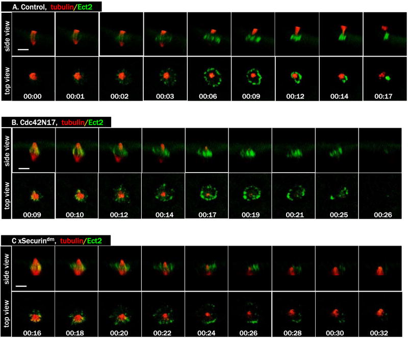
Figure 5. Dynamic Ect2 localization during polar body emission.
A. A series from 4D movie of a control oocyte injected with rhodamine-tubulin (red) and Ect2-3GFP mRNA (green). Ect2 first appears at the central spindle and ends at midbody.
B. A series from 4D movie of an oocyte injected with Cdc42N17, together with rhodamine-tubulin (red) and Ect2-3GFP (green). Ect2 appears at the central spindle but fades away without contraction.
C. A series from 4D movie of an oocyte injected with xSecurindm, together with rhodamine-tubulin (red) and Ect2-3GFP (green). Ect2 signals appear more disorganized (than those in B) and similarly fade quickly.

