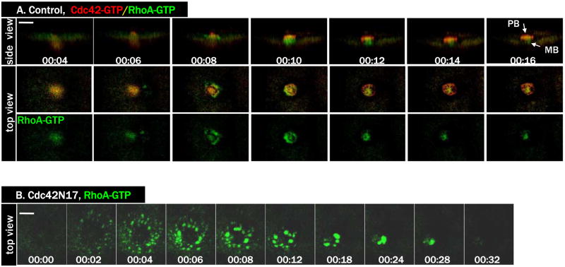
Figure 6. Inhibition of Cdc42 caused hyperactivation of RhoA.
A. A series from 4D movie of a control oocyte exhibiting dynamic and complementary activity zones of RhoA (eGFP-rGBD, green) and Cdc42 (RFP-wGBD, red) during polar body (PB) formation. MB, midbody.
B. A series from 4D movie of a Cdc42N17-injected oocyte with the same two probes as in A. Only eGFP-rGBD signals were shown. Note that RhoA activity zone spreads out in much larger area.

